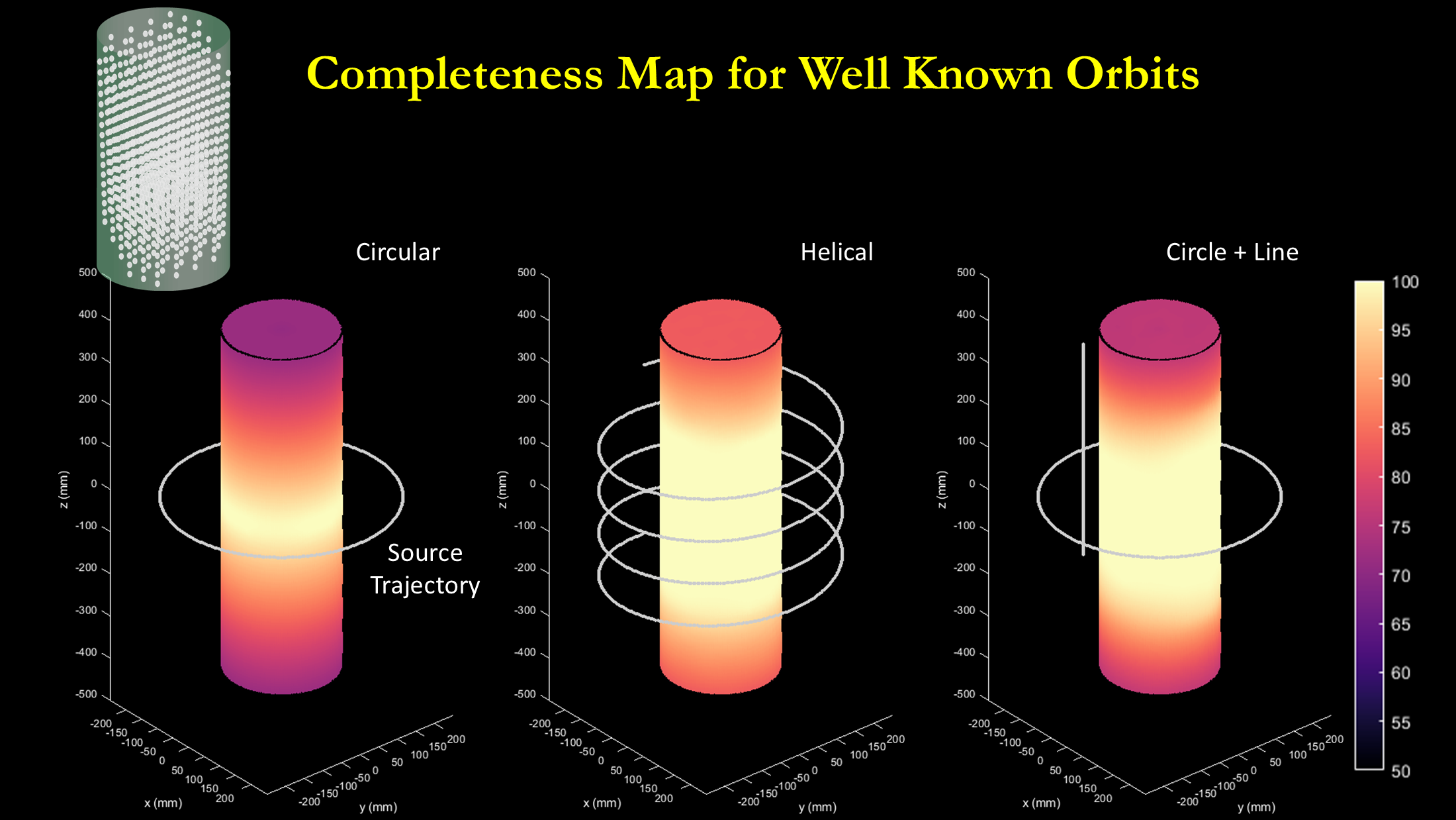
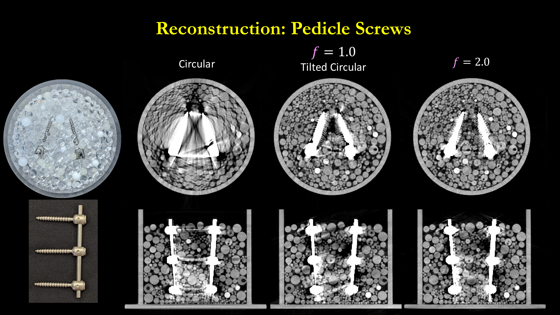
We develop novel acquisition and reconstruction techniques for interventional cone-beam CT, including non-circular orbits for large field-of-view imaging and metal artifact reduction.

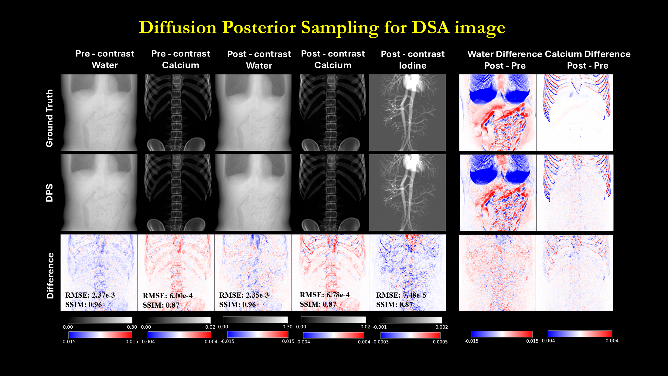
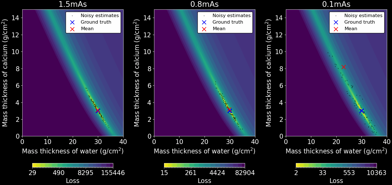
Digital subtraction angiography is sensitive to motion between mask and contrast frames. We develop spectral-temporal processing methods that use energy-resolved measurements to improve vessel visibility, material separation, and robustness to motion in interventional angiography.

We model spectral imaging systems to improve the performance of quantitative spectral imaging tasks. Predictive models for bias, variance, and task-based performance indices are developed to improve spectral detector and system designs.

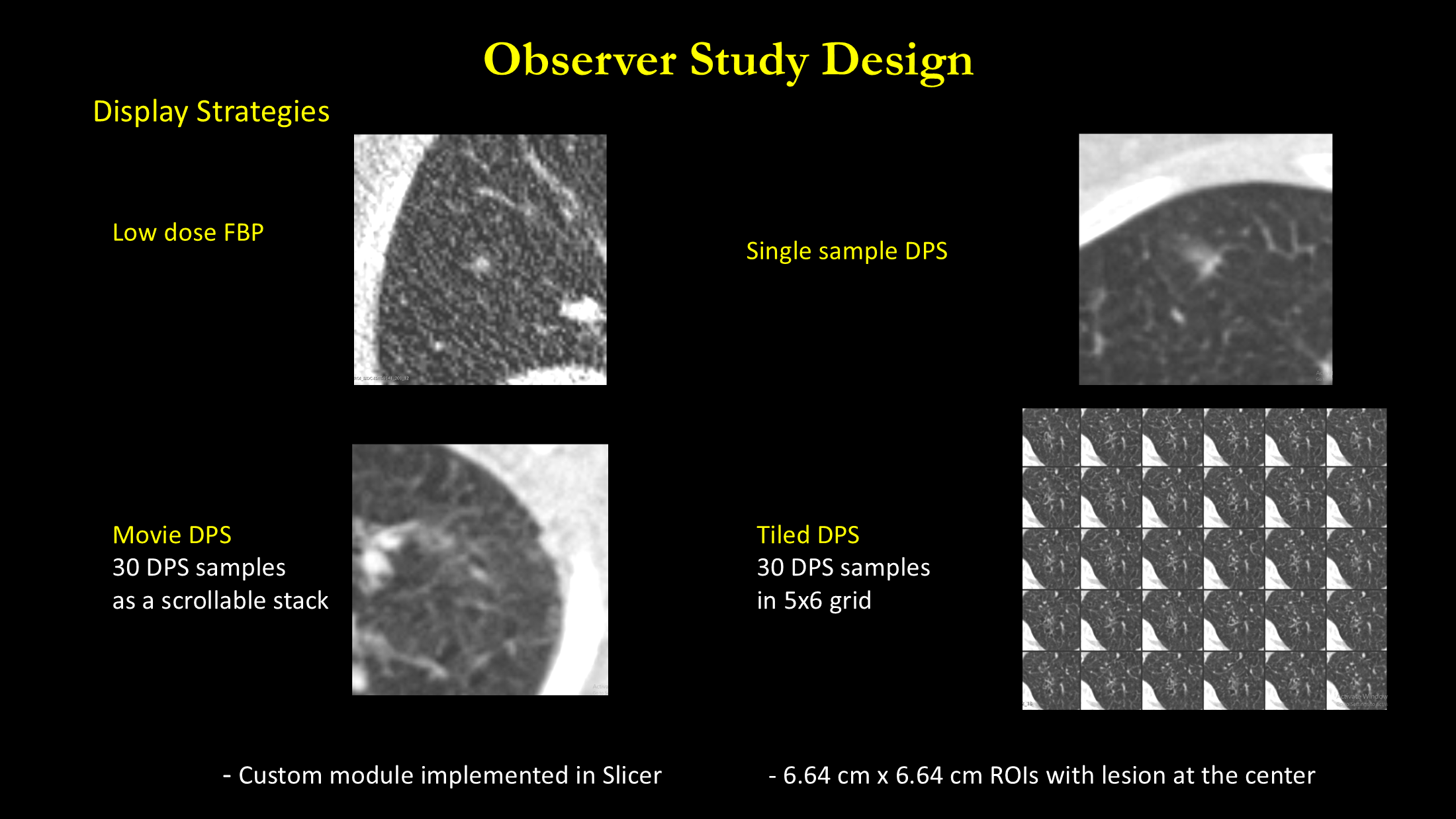
Deep learning reconstruction and post-processing methods are nonlinear and data dependent. We develop analysis methods to evaluate how learned algorithms change image content and diagnostic performance, including how uncertainty and variability can be communicated to radiologists.

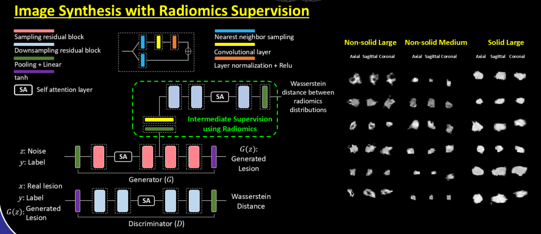
We develop domain-guided generative models for image synthesis. We developed a generative adversarial network with radiomics supervision to encourage synthesized and training samples to have similar radiomic feature distributions. We later extended this concept to radiomics-conditioned diffusion posterior sampling, treating radiomic features as consistency constraints to control the synthesized lesions.


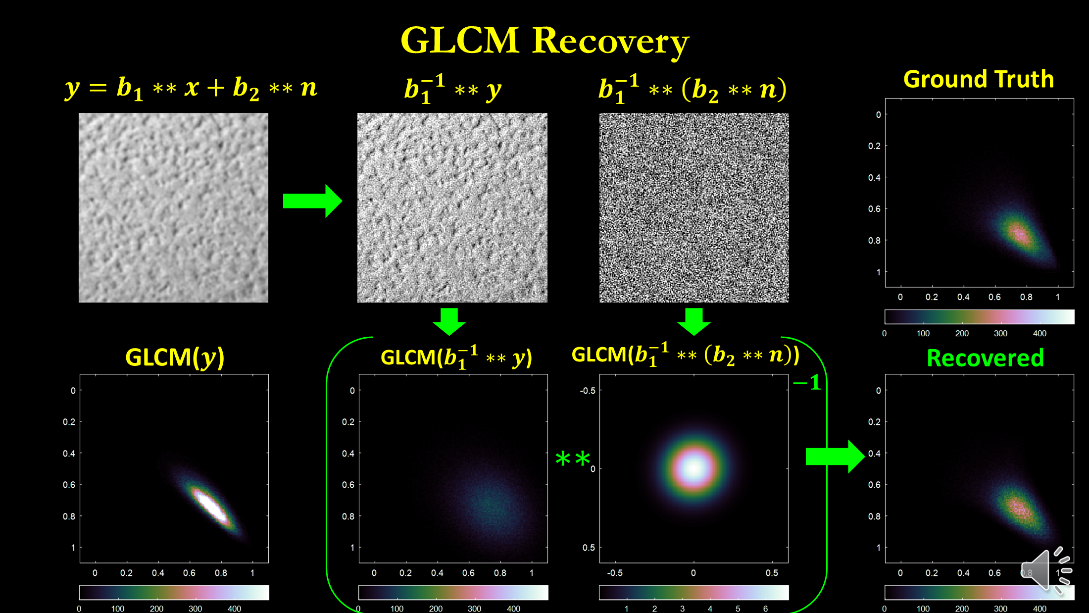
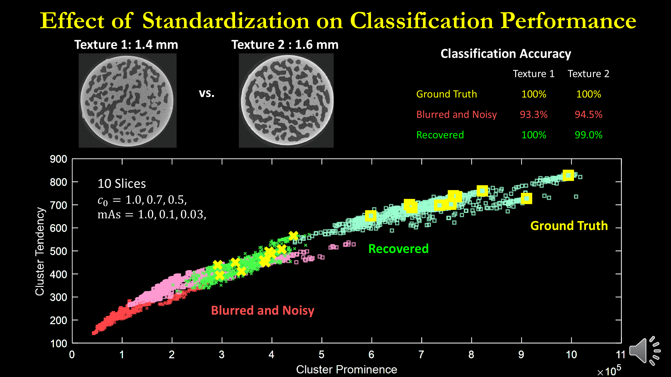
Scanner-induced variability is a major impediment to the clinical translation of radiomics studies. We use prior knowledge of spatial resolution and noise to predict and mitigate scanner-dependent feature variability for robust, reproducible, and generalizable downstream quantitative analysis.

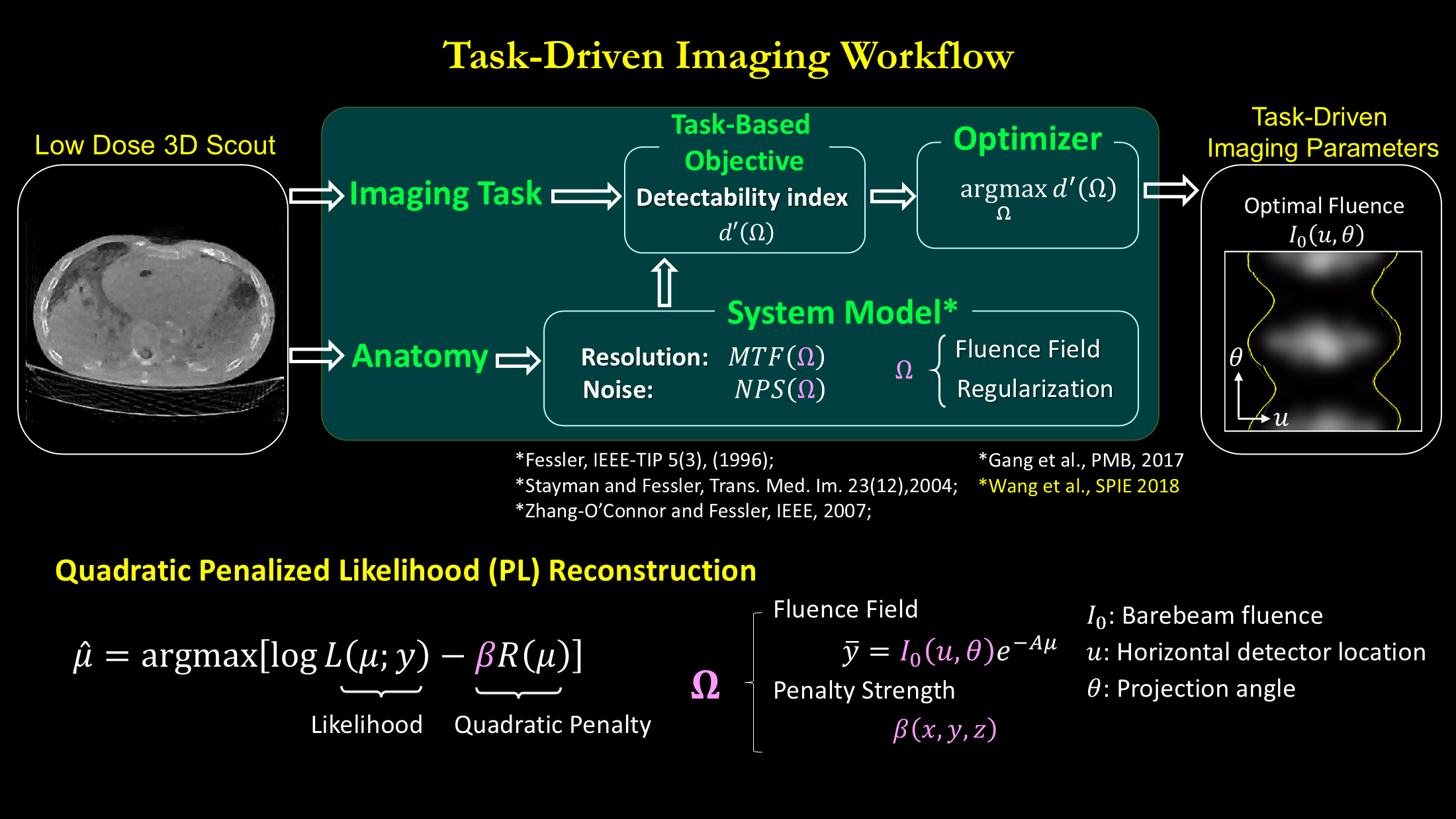
We model the imaging chain from data acquisition through reconstruction and image interpretation. The goal is to use task-based detectability indices to optimize system design and improve imaging performance.

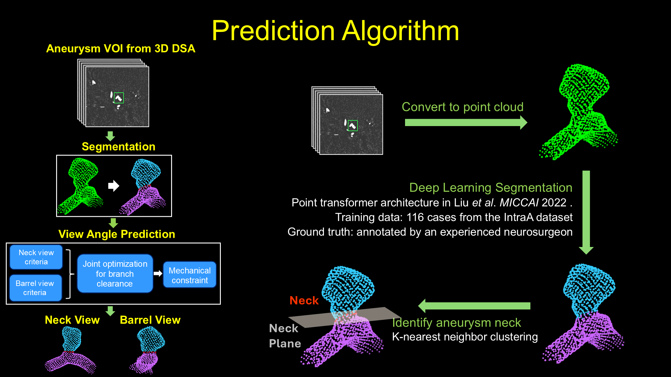
We develop image-analysis and quantitative feature-extraction methods for procedural planning and outcome evaluation. In neuro-interventional applications, this includes automatic working-angle prediction for intracranial aneurysm treatment using anatomical models derived from 3D angiographic imaging.